Data acquisition
Data acquisition#
Issue
Data acquisition is largely carried out with vendored systems. Manufacturers typically keep their software and hardware closed or semi-open at most. As a result, researchers often receive highly processed (e.g., reconstructed) data as ‘raw’ data from the devices. The lack of transparency in the acquisition details and downstream proprietary processing prevents end-to-end reproducible neuroimaging workflows. Reproducibility is endangered, for instance, by heterogeneity in data formats, definition of critical experimental parameters, and technological differences that are translated into the data as spurious, non-biological differences between acquisition devices.
What do we provide
These shortcomings of mostly closed solutions have triggered a growing interest in open-source acquisition hardware and software [Winter et al., 2016]. Here, we provide a brief review of these developments and accompanying solutions aimed at fostering open and collaborative acquisition method development across imaging modalities.
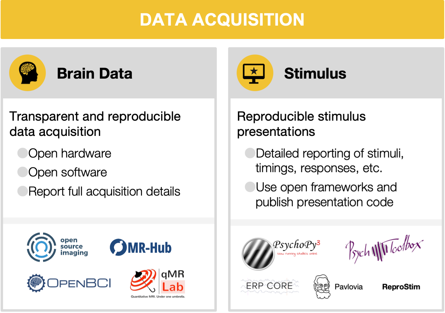
3.1 Brain data acquisition
A common approach advocated by MRI researchers is establishing consensus protocols to standardize data acquisition. One of the flagship applications of this strategy is the Human Connectome Project (HCP) protocol, which achieved this within the confines of a single vendor [Smith et al., 2013]. The HCP acquisition sequences and reconstruction software are compiled for different MRI scanner versions of a single vendor, openly distributed and maintained for fMRI applications [Uğurbil et al., 2013]. However, it is generally difficult to achieve good inter vendor agreement using off the shelf software even for widely used protocols, such as apparent diffusion coefficient and longitudinal relaxation time [Lee et al., 2019, Sasaki et al., 2008]. In addition, not all software options are available from all vendors (for example, compressed sensing [Lustig et al., 2008] and frequency-domain based parallel imaging methods [Breuer et al., 2005, Griswold et al., 2002]). Moreover, even seemingly simple image enhancement protocols, such as image inhomogeneity corrections, are often scarcely documented and validated but can affect inferences drawn from an experiment ([Schmitt and Rieger, 2021]; e.g., [Jellús and Kannengiesser, 2014]). Users typically have access to key parameters of pulse sequences, which are at the center of data acquisition. The exact pulse sequence descriptions are vendor-specific and may even change between software upgrades of a single vendor. This makes it difficult to evaluate multi-center validity of new acquisition methods or to acquire longitudinal data with confidence.
Fortunately, in the last decade, several vendor-neutral data acquisition pulse sequences and reconstruction frameworks have been developed to mitigate this problem: Pulseq [Layton et al., 2017], PyPulseq [Ravi et al., 2019], GammaStar [Cordes et al., 2020], TOPPE [Nielsen and Noll, 2018], ODIN [Jochimsen and von Mengershausen, 2004], and SequenceTree [Magland et al., 2016] (see the resources table). Although these tools vary in vendor compatibility and the flexibility of their acquisition runtime, they enable vendor-neutral deployment of pulse sequences with transparent access to all the details needed. Nevertheless, vendor-neutral raw data (k-space, i.e. the 2D or 3D Fourier space representation of the image) collection is half the battle.
To complete the puzzle of MR image acquisition, interoperable and open-source reconstruction frameworks are essential. Thanks to ISMRM-RD [Inati et al., 2017], a k-space data standard, community-developed reconstruction tools can have a unified way to run advanced reconstruction algorithms against undersampled raw data [Maier et al., 2021]. Some of these tools include Gadgetron [Hansen and Sørensen, 2013], BART [Uecker et al., 2015], MRIReco.Jl [Knopp and Grosser, 2021] (see the resources table for further tools and details). By streamlining these acquisition and reconstruction tools using data standards at multiple levels [Inati et al., 2017, Karakuzu et al., 2021] on a data-driven and container-mediated workflow engine [Di Tommaso et al., 2017], end-to-end reproducible MRI workflows can be developed. A recent study has shown that this approach can significantly reduce inter-vendor variability of quantitative MRI measurements [Karakuzu et al., 2022, Karakuzu et al., 2020]. Given the growing open-source MRI acquisition ecosystem, a variety of end-to-end workflows are possible. Therefore, community-driven validation frameworks have a key importance for interoperable solutions [Tong et al., 2021]. Facilitated by these standards, effective and open communication methods development sets the future direction for reproducible MRI research [Stikov et al., 2019].
In PET, the variety between different scanners is even larger than in MRI. An overview over different scanner types based on their usage for a specific radiotracer targeting the serotonin transporter, namely [11C]DASB, is given in [Nørgaard et al., 2019]. Different PET scanners export images in slightly different data formats with little overlap in the Digital Imaging and Communications in Medicine (DICOM) PET specific tags. As with MRI, reconstruction is vendor/machine specific but open source solutions to image reconstruction are being developed, for instance the OMEGA toolbox [Wettenhovi et al., 2021]. Data acquisition for PET is further complicated by the use of different PET tracers, injection methods, scan duration and scan framing or injected radioactivity dose.
In MEG and EEG, the problem of standardized data acquisition starts even earlier: unlike the common DICOM data format used across vendors in MRI or PET, MEG and EEG manufacturers do not use a common data format, and format specifications are rarely made public. More importantly, equipment implementation significantly differs between vendors, for example with respect to MEG sensor types,(software noise suppression techniques, and EEG amplifiers and electrodes. There have been some efforts on developing open versions of some proprietary tools, for example, the Maxwell filtering for signal space separation by the MNE-python team [Gramfort et al., 2014]. Additionally, initiatives, such as the OpenBCI, offer open EEG hardware and tools for biosensing and brain computer interfacing through continuous community driven development. As we have mentioned, very little is known on how the variability of data acquisition parameters affect downstream comparability of results. The EEGManyLabs project [Pavlov et al., 2021] will provide a comprehensive dataset in this regard, as many labs with different equipment try to replicate the same studies.
Given the large variations across different vendors for all neuroimaging modalities, which often cannot be overcome, it is crucial to report all data acquisition parameters in a comprehensive and standardized manner to make potential differences in data acquisition across studies and sites transparent (for a discussion of reporting guidelines see Section 6.4).
3.2 Stimulus presentation and behavior
Several actively maintained programs for stimulus presentation and response logging are available. Open source software includes PsychoPy [Peirce et al., 2019] in Python and Psychtoolbox [Brainard, 1997, Kleiner et al., 2007, Pelli, 1997] in MATLAB. Both have many users, making it possible to get assistance and perhaps find an already-implemented task protocol (e.g., on Pavlovia for Psychopy). Modality specific resources also exist, for instance the ERP CORE (Compendium of Open Resources and Experiments; Kappenman et al. Kappenman et al. [2021] openly provides optimized paradigms for several widely used ERP components, along with scripts, data processing pipelines, and sample data.
Using open stimuli and presentation software generally increases the likelihood that other researchers can perform replications, because the stimuli and software will be accessible to them. Although desirable, it is not always possible to use fully open stimuli, particularly in the case of commercial movies, audio plays, and image databases. The license of stimuli one wishes to use should always be checked, as should the license one chooses to attach to a dataset when sharing. Stimuli, presentation scripts, behavioral tests and related material should be shared whenever possible (see [DuPre et al., 2019] for a list of datasets sharing naturalistic stimuli and Section 6). To facilitate stimuli feature analysis and exact reproducibility of the experimental paradigms, such projects as ReproNim’s ReproStim [Connolly and Halchenko, 2022] could automate recording and archival of the delivered to audio-video stimulation. When specific stimuli or material can not be released, they should be described as unambiguously as possible and, if possible, providing the source, such as identification number (e.g., a GTIN), and scripts to (re)produce used stimuli from the commercial media.
References on this page
- C1
David H Brainard. The psychophysics toolbox. Spatial Vision, 10(4):433–436, 1997.
- C2
Felix A Breuer, Martin Blaimer, Robin M Heidemann, Matthias F Mueller, Mark A Griswold, and Peter M Jakob. Controlled aliasing in parallel imaging results in higher acceleration (CAIPIRINHA) for multi-slice imaging. Magnetic Resonance in Medicine, 53(3):684–691, March 2005.
- C3
Andy Connolly and Yaroslav Halchenko. ReproNim/reprostim:. March 2022.
- C4
Cristoffer Cordes, Simon Konstandin, David Porter, and Matthias Günther. Portable and platform-independent MR pulse sequence programs. Magnetic Resonance in Medicine, 83(4):1277–1290, April 2020.
- C5
Paolo Di Tommaso, Maria Chatzou, Evan W Floden, Pablo Prieto Barja, Emilio Palumbo, and Cedric Notredame. Nextflow enables reproducible computational workflows. Nature Biotechnology, 35(4):316–319, April 2017.
- C6
Elizabeth DuPre, Michael Hanke, and Jean-Baptiste Poline. Nature abhors a paywall: how open science can realize the potential of naturalistic stimuli. 2019.
- C7
Alexandre Gramfort, Martin Luessi, Eric Larson, Denis A Engemann, Daniel Strohmeier, Christian Brodbeck, Lauri Parkkonen, and Matti S Hämäläinen. MNE software for processing MEG and EEG data. Neuroimage, 86:446–460, February 2014.
- C8
Mark A Griswold, Peter M Jakob, Robin M Heidemann, Mathias Nittka, Vladimir Jellus, Jianmin Wang, Berthold Kiefer, and Axel Haase. Generalized autocalibrating partially parallel acquisitions (GRAPPA). Magnetic Resonance in Medicine, 47(6):1202–1210, 2002.
- C9
Michael Schacht Hansen and Thomas Sangild Sørensen. Gadgetron: an open source framework for medical image reconstruction. Magnetic Resonance in Medicine, 69(6):1768–1776, June 2013.
- C10(1,2)
Souheil J Inati, Joseph D Naegele, Nicholas R Zwart, Vinai Roopchansingh, Martin J Lizak, David C Hansen, Chia-Ying Liu, David Atkinson, Peter Kellman, Sebastian Kozerke, Hui Xue, Adrienne E Campbell-Washburn, Thomas S Sørensen, and Michael S Hansen. ISMRM raw data format: a proposed standard for MRI raw datasets. Magnetic Resonance in Medicine, 77(1):411–421, January 2017.
- C11
V Jellús and S A R Kannengiesser. Adaptive coil combination using a body coil scan as phase reference. Joint Annual Meeting ISMRM-ESMRMB, pages 4406, 2014.
- C12
Thies H Jochimsen and Michael von Mengershausen. ODIN: object-oriented development interface for NMR. Journal of Magnetic Resonance, 170(1):67–78, 2004.
- C13
Emily S Kappenman, Jaclyn L Farrens, Wendy Zhang, Andrew X Stewart, and Steven J Luck. ERP CORE: an open resource for human event-related potential research. Neuroimage, 225:117465, January 2021.
- C14
Agah Karakuzu, Stefan Appelhoff, Tibor Auer, Mathieu Boudreau, Franklin Feingold, Ali R Khan, Alberto Lazari, Christopher Markiewicz, Martjin j Mulder, Christophe Phillips, Taylor Salo, Nikola Stikov, Kirstie Whitaker, and Gilles Hollander. qMRI-BIDS: an extension to the brain imaging data structure for quantitative magnetic resonance imaging data. medRxiv, pages 2021.10.22.21265382, October 2021.
- C15
Agah Karakuzu, Labonny Biswas, Julien Cohen‐Adad, and Nikola Stikov. Vendor‐neutral sequences and fully transparent workflows improve inter‐vendor reproducibility of quantitative mri. Magnetic Resonance in Medicine, 88(3):1212–1228, 2022.
- C16
Agah Karakuzu, Mathieu Boudreau, Tanguy Duval, Tommy Boshkovski, Ilana Leppert, Jean-François Cabana, Ian Gagnon, Pascale Beliveau, G Pike, Julien Cohen-Adad, and Nikola Stikov. qMRLab: quantitative MRI analysis, under one umbrella. Journal of Open Source Software, 5(53):2343, September 2020.
- C17
M Kleiner, D Brainard, and D Pelli. What's new in psychtoolbox-3? Perception, 2007.
- C18
Tobias Knopp and Mirco Grosser. MRIReco.jl: an MRI reconstruction framework written in julia. Magnetic Resonance in Medicine, 86(3):1633–1646, September 2021.
- C19
Kelvin J Layton, Stefan Kroboth, Feng Jia, Sebastian Littin, Huijun Yu, Jochen Leupold, Jon-Fredrik Nielsen, Tony Stöcker, and Maxim Zaitsev. Pulseq: a rapid and hardware-independent pulse sequence prototyping framework. Magnetic Resonance in Medicine, 77(4):1544–1552, April 2017.
- C20
Yoojin Lee, Martina F Callaghan, Julio Acosta-Cabronero, Antoine Lutti, and Zoltan Nagy. Establishing intra- and inter-vendor reproducibility of T relaxation time measurements with 3T MRI. Magnetic Resonance in Medicine, 81(1):454–465, January 2019.
- C21
M Lustig, D L Donoho, J M Santos, and J M Pauly. Compressed sensing MRI. IEEE Signal Processing Magazine, 25(2):72–82, March 2008.
- C22
Jeremy F Magland, Cheng Li, Michael C Langham, and Felix W Wehrli. Pulse sequence programming in a dynamic visual environment: SequenceTree. Magnetic Resonance in Medicine, 75(1):257–265, January 2016.
- C23
Oliver Maier, Steven Hubert Baete, Alexander Fyrdahl, Kerstin Hammernik, Seb Harrevelt, Lars Kasper, Agah Karakuzu, Michael Loecher, Franz Patzig, Ye Tian, Ke Wang, Daniel Gallichan, Martin Uecker, and Florian Knoll. CG-SENSE revisited: results from the first ISMRM reproducibility challenge. Magnetic Resonance in Medicine, 85(4):1821–1839, April 2021.
- C24
Jon-Fredrik Nielsen and Douglas C Noll. TOPPE: a framework for rapid prototyping of MR pulse sequences. Magnetic Resonance in Medicine, 79(6):3128–3134, June 2018.
- C25
Martin Nørgaard, Melanie Ganz, Claus Svarer, Ling Feng, Masanori Ichise, Rupert Lanzenberger, Mark Lubberink, Ramin V Parsey, Marios Politis, Eugenii A Rabiner, Mark Slifstein, Vesna Sossi, Tetsuya Suhara, Peter S Talbot, Federico Turkheimer, Stephen C Strother, and Gitte M Knudsen. Cerebral serotonin transporter measurements with [11C]DASB: a review on acquisition and preprocessing across 21 PET centres. Journal of Cerebral Blood Flow & Metabolism, 39(2):210–222, 2019.
- C26
Yuri G Pavlov, Nika Adamian, Stefan Appelhoff, Mahnaz Arvaneh, Christopher S Y Benwell, Christian Beste, Amy R Bland, Daniel E Bradford, Florian Bublatzky, Niko A Busch, Peter E Clayson, Damian Cruse, Artur Czeszumski, Anna Dreber, Guillaume Dumas, Benedikt Ehinger, Giorgio Ganis, Xun He, José A Hinojosa, Christoph Huber-Huber, Michael Inzlicht, Bradley N Jack, Magnus Johannesson, Rhiannon Jones, Evgenii Kalenkovich, Laura Kaltwasser, Hamid Karimi-Rouzbahani, Andreas Keil, Peter König, Layla Kouara, Louisa Kulke, Cecile D Ladouceur, Nicolas Langer, Heinrich R Liesefeld, David Luque, Annmarie MacNamara, Liad Mudrik, Muthuraman Muthuraman, Lauren B Neal, Gustav Nilsonne, Guiomar Niso, Sebastian Ocklenburg, Robert Oostenveld, Cyril R Pernet, Gilles Pourtois, Manuela Ruzzoli, Sarah M Sass, Alexandre Schaefer, Magdalena Senderecka, Joel S Snyder, Christian K Tamnes, Emmanuelle Tognoli, Marieke K van Vugt, Edelyn Verona, Robin Vloeberghs, Dominik Welke, Jan R Wessel, Ilya Zakharov, and Faisal Mushtaq. #EEGManyLabs: investigating the replicability of influential EEG experiments. Cortex, 144:213–229, April 2021.
- C27
Jonathan Peirce, Jeremy R Gray, Sol Simpson, Michael MacAskill, Richard Höchenberger, Hiroyuki Sogo, Erik Kastman, and Jonas Kristoffer Lindeløv. PsychoPy2: experiments in behavior made easy. Behavior Research Methods, 51(1):195–203, February 2019.
- C28
D G Pelli. The VideoToolbox software for visual psychophysics: transforming numbers into movies. Spatial Vision, 10(4):437–442, 1997.
- C29
Keerthi Ravi, Sairam Geethanath, and John Vaughan. PyPulseq: a python package for MRI pulse sequence design. Journal of Open Source Software, 4(42):1725, October 2019.
- C30
Makoto Sasaki, Kei Yamada, Yoshiyuki Watanabe, Mieko Matsui, Masahiro Ida, Shunrou Fujiwara, Eri Shibata, and Acute Stroke Imaging Standardization Group-Japan (ASIST-Japan) Investigators. Variability in absolute apparent diffusion coefficient values across different platforms may be substantial: a multivendor, multi-institutional comparison study. Radiology, 249(2):624–630, November 2008.
- C31
Tina Schmitt and Jochem W Rieger. Recommendations of choice of head coil and prescan normalize filter depend on region of interest and task. Frontiers in Neuroscience, 2021.
- C32
Stephen M Smith, Christian F Beckmann, Jesper Andersson, Edward J Auerbach, Janine Bijsterbosch, Gwenaëlle Douaud, Eugene Duff, David A Feinberg, Ludovica Griffanti, Michael P Harms, Michael Kelly, Timothy Laumann, Karla L Miller, Steen Moeller, Steve Petersen, Jonathan Power, Gholamreza Salimi-Khorshidi, Abraham Z Snyder, An T Vu, Mark W Woolrich, Junqian Xu, Essa Yacoub, Kamil Uğurbil, David C Van Essen, Matthew F Glasser, and WU-Minn HCP Consortium. Resting-state fMRI in the human connectome project. Neuroimage, 80:144–168, October 2013.
- C33
Nikola Stikov, Joshua D Trzasko, and Matt A Bernstein. Reproducibility and the future of MRI research. Magnetic Resonance in Medicine, 82(6):1981–1983, December 2019.
- C34
Gehua Tong, Andreia S Gaspar, Enlin Qian, Keerthi Sravan Ravi, John Thomas Vaughan, Jr, Rita G Nunes, and Sairam Geethanath. A framework for validating open-source pulse sequences. Magnetic Resonance Imaging, 87:7–18, November 2021.
- C35
Martin Uecker, Frank Ong, Jonathan I Tamir, Dara Bahri, Patrick Virtue, Joseph Y Cheng, Tao Zhang, and Michael Lustig. Berkeley advanced reconstruction toolbox (BART). Proc. of. the International Society for Magnetic Resonance in Medicine (ISMRM), October 2015.
- C36
Kamil Uğurbil, Junqian Xu, Edward J Auerbach, Steen Moeller, An T Vu, Julio M Duarte-Carvajalino, Christophe Lenglet, Xiaoping Wu, Sebastian Schmitter, Pierre Francois Van de Moortele, John Strupp, Guillermo Sapiro, Federico De Martino, Dingxin Wang, Noam Harel, Michael Garwood, Liyong Chen, David A Feinberg, Stephen M Smith, Karla L Miller, Stamatios N Sotiropoulos, Saad Jbabdi, Jesper L R Andersson, Timothy E J Behrens, Matthew F Glasser, David C Van Essen, Essa Yacoub, and WU-Minn HCP Consortium. Pushing spatial and temporal resolution for functional and diffusion MRI in the human connectome project. Neuroimage, 80:80–104, October 2013.
- C37
V-V Wettenhovi, M Vauhkonen, and V Kolehmainen. OMEGA: open-source emission tomography software. Physics in Medicine and Biology, 2021.
- C38
Lukas Winter, H Haopeng, Antonia Barghoorn, Werner Hoffmann, Stefan Hetzer, Simone Winkler, and Others. Open source imaging initiative. Proc. of. the International Society for Magnetic Resonance in Medicine (ISMRM), 2016.
- 1
Sources: Icons from the Noun Project: Brain by parkjisun; Computer Screen by Icon Solid (adapted with a star); Logos: used with permission by the copyright holders.